Cells are the basic structural and functional units of living organisms. Each cell is composed of various organelles that are responsible for specific functions within the cell. In this article, we will explore the different cell organelles, their structures, and their functions.
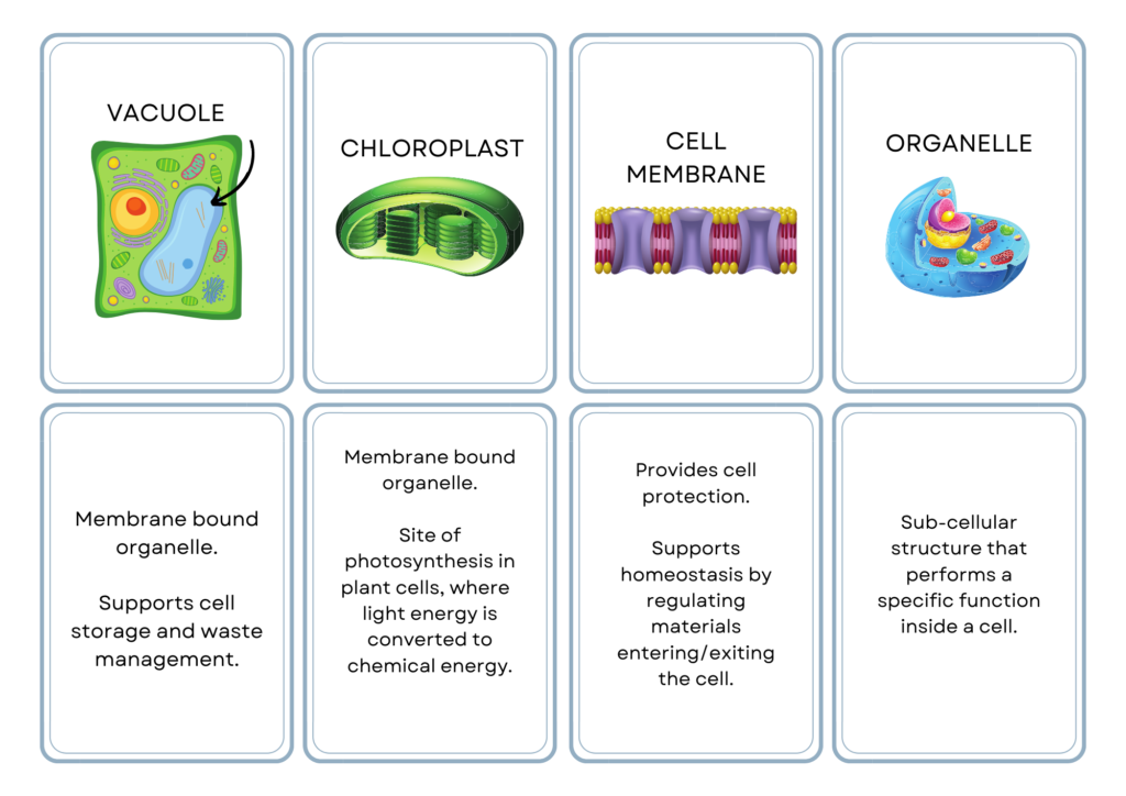
A cell organelle is a specialized structure within a cell that performs a specific function. These organelles are found in eukaryotic cells, which are cells that have a nucleus and other membrane-bound organelles. Prokaryotic cells, such as bacteria, do not have membrane-bound organelles.
Cell Organelles – Structure and Functions
1. Nucleus
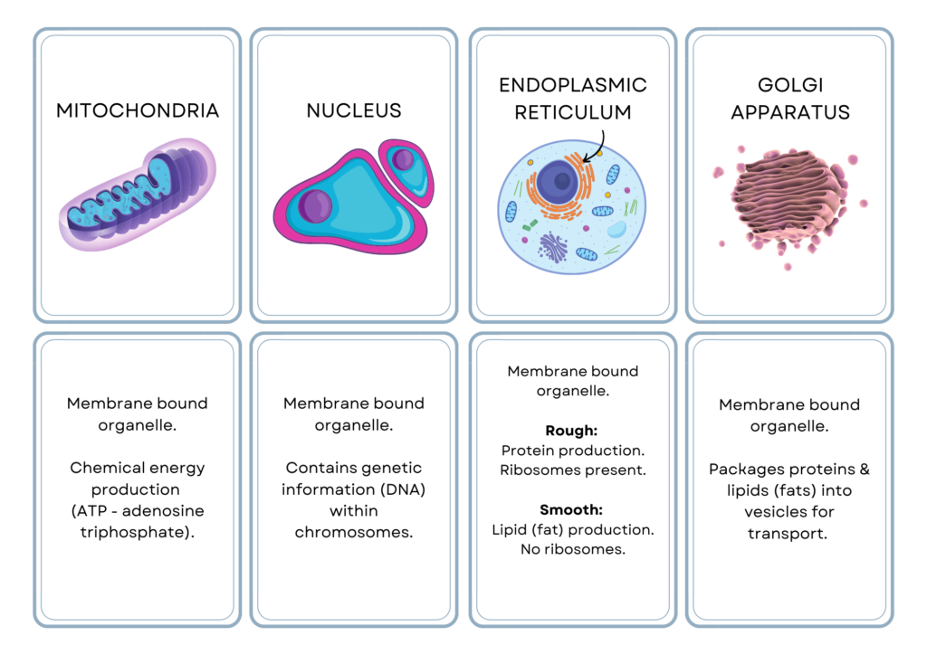
The nucleus is a critical organelle that is present in most eukaryotic cells. It is responsible for controlling cellular activities by regulating gene expression, DNA replication, and RNA synthesis. The structure of the nucleus is quite complex and consists of several components, including the nuclear envelope, chromatin, nucleolus, and nuclear pores. In this article, we will discuss the structure and functions of the nucleus in detail.
Structure of the Nucleus
- The nucleus is a spherical or oval-shaped organelle that is usually located in the center of the cell.
- It is surrounded by a double-layered membrane called the nuclear envelope.
- The nuclear envelope separates the nucleus from the cytoplasm and has tiny openings called nuclear pores that allow the passage of molecules between the nucleus and the cytoplasm.
- The nuclear envelope consists of an outer membrane and an inner membrane. The space between these two membranes is called the perinuclear space.
- The inner membrane is lined by a network of intermediate filaments called the nuclear lamina, which provides structural support to the nucleus.
- The genetic material of the cell is stored in the nucleus in the form of chromatin.
- Chromatin is made up of DNA and proteins, and it exists in two forms: euchromatin and heterochromatin.
- Euchromatin is the less condensed and more active form of chromatin, whereas heterochromatin is the more condensed and less active form.
- The nucleolus is a dense, spherical structure present within the nucleus. It is involved in the synthesis and assembly of ribosomal subunits.
- The nucleolus is made up of ribosomal DNA (rDNA) and several proteins.
Functions of the Nucleus
The nucleus is responsible for controlling cellular activities by regulating gene expression. It contains the genetic material of the cell, which is responsible for determining the traits and characteristics of an organism. The following are the functions of the nucleus:
- DNA Replication: The nucleus is responsible for the replication of DNA, which is necessary for the growth and division of cells. During DNA replication, the double-stranded DNA molecule is unwound, and each strand serves as a template for the synthesis of a new complementary strand.
- RNA Synthesis: The nucleus is responsible for the synthesis of RNA, which is necessary for the synthesis of proteins. RNA is synthesized from DNA by a process called transcription. During transcription, the DNA sequence is copied into a complementary RNA sequence by RNA polymerase.
- Gene Expression: The nucleus is responsible for regulating gene expression, which determines the traits and characteristics of an organism. Gene expression is regulated by a complex network of signaling pathways and gene regulatory proteins.
- Ribosome Assembly: The nucleolus is responsible for the assembly of ribosomal subunits, which are necessary for the synthesis of proteins. Ribosomal subunits are assembled from rRNA and proteins.
2. Mitochondria
Mitochondria are membrane-bound organelles found in the cytoplasm of eukaryotic cells. These organelles are known as the “powerhouses” of the cell because they are responsible for producing most of the energy that the cell needs to carry out its functions. In this article, we will explore the structure and functions of mitochondria in more detail.
Structure of Mitochondria:
- Mitochondria are generally oval or sausage-shaped organelles, with a double membrane structure.
- The outer membrane is smooth, while the inner membrane is highly folded, forming structures called cristae.
- The cristae greatly increase the surface area of the inner membrane, providing more space for the enzymes involved in cellular respiration.
- The inner membrane also contains proteins that are involved in the electron transport chain, a process that produces ATP, the energy currency of the cell.
- The space between the two membranes is called the intermembrane space, while the fluid-filled space inside the inner membrane is called the mitochondrial matrix.
- The matrix contains enzymes, ribosomes, and mitochondrial DNA, which are involved in the synthesis of proteins required for the functioning of the mitochondria.
Functions of Mitochondria:
- The primary function of mitochondria is to produce energy in the form of ATP through a process called cellular respiration.
- This process involves the breakdown of glucose and other organic molecules in the presence of oxygen to produce ATP.
- Mitochondria are therefore most abundant in cells that require a lot of energy, such as muscle cells, liver cells, and nerve cells.
- Apart from energy production, mitochondria also play a role in regulating calcium ion levels in the cell, which is important for various cellular processes.
- They also play a role in programmed cell death or apoptosis, as well as in the synthesis of certain hormones and other cellular signaling molecules.
- In addition, mitochondria are believed to have evolved from free-living bacteria that were engulfed by primitive eukaryotic cells.
- This is supported by the fact that mitochondria have their own DNA, which is separate from the DNA in the nucleus of the cell. This DNA contains genes that are essential for the functioning of the mitochondria.
3. Endoplasmic Reticulum
The endoplasmic reticulum (ER) is an organelle that is present in eukaryotic cells. It is a network of flattened sacs, tubes, and cisternae that are interconnected and extend throughout the cytoplasm. The ER is divided into two types, the rough endoplasmic reticulum (RER) and the smooth endoplasmic reticulum (SER), based on their structure and functions.
Structure:
- The RER has ribosomes attached to its surface, giving it a rough appearance.
- The ribosomes on the RER are responsible for the synthesis of proteins that are destined for secretion or membrane insertion.
- The SER lacks ribosomes, and its surface is smooth.
- The SER is involved in lipid synthesis, carbohydrate metabolism, and detoxification reactions.
Functions:
- The RER plays a crucial role in protein synthesis, folding, and modification.
- The proteins synthesized by the ribosomes on the RER are transported into the lumen of the ER, where they undergo folding, modification, and sorting. The RER also plays a vital role in the quality control of newly synthesized proteins.
- Misfolded proteins are recognized by chaperones, which either refold them or target them for degradation.
- The SER has various functions, depending on the cell type. In liver cells, the SER is responsible for the detoxification of drugs and toxins.
- It accomplishes this by modifying the lipophilic molecules into more water-soluble forms that can be excreted in the urine.
- The SER in muscle cells plays a role in calcium storage and release, which is essential for muscle contraction.
- In adipose cells, the SER is involved in lipid synthesis and metabolism.
- The ER also plays a critical role in the maintenance of calcium homeostasis.
- The ER lumen is a calcium store, and the calcium concentration is tightly regulated by pumps and channels that transport calcium ions in and out of the ER.
- Calcium release from the ER triggers a variety of cellular processes, including muscle contraction and cell signaling.
4. Golgi Apparatus
The Golgi apparatus, also known as the Golgi complex or Golgi body, is an organelle found in eukaryotic cells. It plays a crucial role in the processing and sorting of proteins and lipids and is involved in the transport of these molecules to their final destinations within the cell or outside of it.
Structure
- The Golgi apparatus is composed of stacks of flattened, membrane-bound sacs called cisternae.
- These stacks are arranged in a characteristic “pancake” shape and are often compared to a stack of plates.
- The cisternae are surrounded by vesicles that transport molecules in and out of the Golgi apparatus.
- The Golgi apparatus is divided into three regions: the cis-Golgi, medial-Golgi, and trans-Golgi.
- The cis-Golgi is closest to the endoplasmic reticulum (ER), and receives newly synthesized proteins and lipids from the ER.
- The medial Golgi is located in the middle of the stack and is involved in the processing and modification of these molecules.
- The trans-Golgi is located at the opposite end of the stack and is responsible for the final sorting and packaging of the molecules.
Functions
The Golgi apparatus has several important functions, including:
- Protein Processing and Sorting: The Golgi apparatus is responsible for the modification and sorting of proteins that are synthesized in the endoplasmic reticulum. The proteins are transported to the cis-Golgi, where they are modified by the addition or removal of carbohydrate groups or other molecules. These modifications can change the function of the protein or target it to a specific location within the cell or outside of it.
- Lipid Processing and Sorting: In addition to proteins, the Golgi apparatus is also involved in the processing and sorting of lipids. The lipids are transported to the cis-Golgi, where they are modified and sorted into vesicles for transport to their final destination.
- Vesicle Formation: The Golgi apparatus is involved in the formation of vesicles that transport molecules to their final destination. Vesicles bud off from the Golgi apparatus and are transported to the plasma membrane, where they fuse with the membrane and release their contents outside of the cell.
- Glycosylation: The Golgi apparatus is involved in the process of glycosylation, which is the addition of carbohydrate groups to proteins and lipids. This modification can change the function of the molecule or target it to a specific location within the cell or outside of it.
5. Lysosomes
Lysosomes are organelles found in animal cells that are involved in the breakdown and recycling of cellular waste and foreign material. In this article, we will explore the structure and functions of lysosomes in detail.
Structure
- Lysosomes are membrane-bound organelles that contain digestive enzymes. They are formed by the Golgi apparatus, which packages the enzymes into vesicles that are then transported to the lysosome.
- The membrane of the lysosome is composed of lipids and proteins, and it contains pumps and channels that transport materials in and out of the organelle.
- The enzymes within the lysosome are acid hydrolases, which require an acidic environment to function.
Functions
Lysosomes have several important functions, including:
- Digestion: Lysosomes are responsible for the breakdown of cellular waste and foreign material. They fuse with incoming vesicles or phagosomes, which contain material to be broken down, and release their enzymes into the vesicle or phagosome. The enzymes then break down the material into its basic components, which can be recycled or excreted by the cell.
- Autophagy: Lysosomes are also involved in a process called autophagy, which is the breakdown of cellular components that are no longer needed or are damaged. The lysosome fuses with the autophagosome, which contains the material to be broken down, and releases its enzymes into the autophagosome. The enzymes then break down the material, allowing its components to be recycled or excreted.
- Immunity: Lysosomes are involved in the immune response of the cell. They can fuse with phagosomes containing foreign material, such as bacteria or viruses, and break them down. This helps to protect the cell from infection and disease.
- Signaling: Lysosomes are also involved in signaling pathways within the cell. They can release enzymes and other molecules into the cytoplasm, which can activate or inhibit various cellular processes.
6. Peroxisomes
Peroxisomes are small, single membrane-bound organelles found in most eukaryotic cells. These organelles are involved in a variety of metabolic functions, including fatty acid metabolism and the breakdown of toxic substances in cells. In this article, we will discuss the structure and functions of peroxisomes in more detail.
Structure of Peroxisomes
- Peroxisomes are small, spherical or oval-shaped organelles that range in size from 0.1 to 1.0 micrometers in diameter.
- They are surrounded by a single membrane that is similar in composition to the plasma membrane. The interior of the peroxisome is filled with a liquid matrix that contains a variety of enzymes.
- Peroxisomes contain several types of enzymes that are involved in a variety of metabolic processes.
- One of the most important enzymes found in peroxisomes is catalase. Catalase is involved in the breakdown of hydrogen peroxide, a toxic byproduct of many cellular reactions.
- Peroxisomes also contain other enzymes, such as oxidases, which are involved in the breakdown of fatty acids and other substances.
Functions of Peroxisomes
- The main function of peroxisomes is the breakdown of fatty acids. Peroxisomes contain several enzymes that are involved in this process, including acyl-CoA oxidase, which breaks down fatty acids into acetyl-CoA. This molecule can then be used by the cell to produce energy.
- In addition to fatty acid metabolism, peroxisomes are also involved in the breakdown of other substances. For example, peroxisomes contain enzymes that are involved in the breakdown of purines and polyamines, which are important components of nucleic acids.
- Another important function of peroxisomes is the detoxification of harmful substances in cells. Peroxisomes contain several enzymes, including catalase, that are involved in the breakdown of hydrogen peroxide and other toxic substances.
- Peroxisomes are also involved in the biosynthesis of certain molecules. For example, peroxisomes are involved in the biosynthesis of plasmalogens, which are important components of cell membranes.
7. Plasma Membranes
The plasma membrane is a thin, flexible barrier that surrounds and encloses the contents of the cell. It is composed of a lipid bilayer that separates the interior of the cell from the outside environment. In this article, we will explore the structure and functions of the plasma membrane.
Structure
- The plasma membrane is composed of a lipid bilayer, which is made up of two layers of phospholipid molecules.
- Each phospholipid molecule consists of a hydrophilic (water-loving) head and a hydrophobic (water-fearing) tail.
- The hydrophilic heads face outward, while the hydrophobic tails face inward, forming a barrier that separates the inside of the cell from the outside environment.
- The lipid bilayer also contains proteins that are embedded in the membrane.
- These proteins have a variety of functions, such as serving as channels or pumps for molecules to cross the membrane, acting as receptors for signaling molecules, and providing structural support to the membrane.
- In addition to phospholipids and proteins, the plasma membrane also contains carbohydrates in the form of glycoproteins and glycolipids. These carbohydrates serve as markers that allow the cell to identify and communicate with other cells.
Functions
The plasma membrane has several important functions, including:
- Selective Permeability: The plasma membrane is selectively permeable, which means that it allows certain substances to pass through while preventing others from entering or leaving the cell. This is essential for maintaining the proper internal environment of the cell and for regulating the movement of molecules in and out of the cell.
- Cell Signaling: The plasma membrane plays an important role in cell signaling. The proteins embedded in the membrane can act as receptors for signaling molecules, such as hormones or neurotransmitters. When a signaling molecule binds to its receptor, it can trigger a cascade of chemical reactions within the cell, leading to a specific cellular response.
- Cell Adhesion: The plasma membrane also plays a role in cell adhesion. Cells can adhere to each other and to extracellular structures, such as the extracellular matrix, through specialized proteins that are embedded in the plasma membrane.
8. Cytoskeleton
The cytoskeleton is a network of protein filaments that give shape and support to cells. It is made up of three types of filaments: microfilaments, intermediate filaments, and microtubules. The cytoskeleton has a range of functions, from maintaining cell shape to allowing cells to move and divide.
Structure
- Microfilaments are the smallest filaments of the cytoskeleton, made up of actin protein monomers that form long chains.
- They are about 7 nm in diameter and are responsible for cell movement, contraction, and cell shape maintenance.
- Intermediate filaments are made up of a variety of protein monomers, such as keratin, and are about 10 nm in diameter.
- They are responsible for maintaining the structural integrity of cells and providing mechanical strength to cells.
- Microtubules are the largest filaments of the cytoskeleton, made up of tubulin protein dimers that form long tubes.
- They are about 25 nm in diameter and are responsible for cell division, cell shape maintenance, and the movement of organelles and vesicles within cells.
Functions
The cytoskeleton has a variety of functions, including:
- Maintaining cell shape: The cytoskeleton provides structural support to cells, allowing them to maintain their shape.
- Cell movement: The cytoskeleton plays a key role in cell movement. Microfilaments and microtubules work together to generate the force required for cell movement. For example, during cell division, microtubules form a spindle that separates the chromosomes, while microfilaments pull the cell membrane inward to divide the cell.
- Intracellular transport: The cytoskeleton plays a crucial role in the transport of organelles and vesicles within cells. Microtubules act as tracks that allow organelles to move from one part of the cell to another.
- Cell signaling: The cytoskeleton is involved in cell signaling, allowing cells to sense and respond to changes in their environment. For example, microfilaments can contract in response to a chemical signal, changing the shape of the cell and allowing it to move.
- Cell division: The cytoskeleton is critical for cell division. Microtubules form the spindle that separates the chromosomes during cell division, while microfilaments pull the cell membrane inward to divide the cell.
9. Vacuoles
Vacuoles are organelles found in plant and fungal cells that are involved in a variety of functions, including storage, waste disposal, and regulation of cell turgor pressure. In this article, we will explore the structure and functions of vacuoles in detail.
Structure:
- Vacuoles are large, membrane-bound organelles that occupy a significant portion of the volume of plant and fungal cells. They are surrounded by a single membrane called the tonoplast, which separates the contents of the vacuole from the rest of the cell.
- The interior of the vacuole is filled with a fluid called the cell sap, which is composed of water, ions, and various organic molecules. The cell sap can be acidic or basic depending on the type of vacuole and its function.
- In addition to the cell sap, vacuoles can contain a variety of substances, such as pigments, starch, proteins, and waste products. Some specialized vacuoles, such as contractile vacuoles found in certain freshwater protists, can also contain specific ions and molecules that are involved in osmoregulation.
Functions
Vacuoles have several important functions, including:
- Storage: Vacuoles are used by the plant and fungal cells as storage organelles. They can store a variety of substances, such as sugars, amino acids, lipids, and ions. In addition, vacuoles can store pigments that are involved in flower coloration and other plant-related functions.
- Waste Disposal: Vacuoles are also involved in waste disposal. They can accumulate and isolate harmful substances, such as metabolic waste products and toxic compounds, and prevent them from damaging the rest of the cell.
- Osmoregulation: In some organisms, such as freshwater protists and plants, vacuoles are involved in osmoregulation, which is the regulation of water and ion balance within the cell. Contractile vacuoles found in certain freshwater protists, for example, are involved in pumping the excess water out of the cell to prevent it from bursting.\
- Regulation of Turgor Pressure: Vacuoles are also involved in regulating turgor pressure within plant cells. Turgor pressure is the pressure that water exerts on the cell wall, which helps maintain the rigidity and shape of the cell. By controlling the movement of water in and out of the vacuole, plants can regulate turgor pressure and maintain their shape and structure.
Frequently Asked Questions (FAQs)
What are cell organelles?
Cell organelles are specialized structures within a cell that carry out specific functions. They include the nucleus, mitochondria, ribosomes, endoplasmic reticulum, Golgi apparatus, lysosomes, peroxisomes, vacuoles, and cytoskeleton.
Can cell organelles be found in both animal and plant cells?
Yes, most cell organelles can be found in both animal and plant cells. However, plant cells also have additional organelles structure such as chloroplasts and a cell wall.
Can cells function properly without organelles?
No, cells cannot function properly without organelles. Organelles are essential for carrying out specialized functions within a cell, such as energy production, protein synthesis, and waste removal.
How do organelles communicate with each other within a cell?
Organelles communicate with each other through a network of channels called the endomembrane system. This system includes the endoplasmic reticulum, Golgi apparatus, and vesicles, which transport molecules between organelles.
Can cell organelles be targeted by drugs?
Yes, some drugs target specific organelles within cells. For example, chemotherapy drugs target the DNA in the nucleus, while antibiotics target the ribosomes responsible for protein synthesis.
Can cell organelles be damaged by environmental factors?
Yes, the organelles can be damaged by environmental factors such as toxins, radiation, and temperature extremes. This can disrupt normal cell functions and lead to cell death or disease.
Summary of Cell Organelles
In conclusion, cells are composed of various organelles that are responsible for specific functions within the cell. Each organelle has a unique structure and function that is essential to the proper functioning of the cell.
Understanding the structure and function of these organelles is important for understanding how cells work and how they can be affected by disease and other factors.