Myoglobin (Mb) is a small globular protein that binds and stores oxygen in cardiac and skeletal muscle cells. It is the main oxygen‑binding protein found in muscle and acts as a local oxygen reservoir, helping muscle fibers continue working when blood oxygen briefly falls during exercise or stress.
Structurally, myoglobin is a monomeric heme protein with a single polypeptide chain and a well‑defined tertiary structure, making it a classic textbook example used to explain the structure and function of proteins.

Because of its high affinity for oxygen, myoglobin plays a key role in aerobic metabolism and is often discussed along with hemoglobin in topics like “myoglobin structure and function” and “difference between myoglobin and hemoglobin structure.”
In exams, myoglobin is often asked about as “What is myoglobin?” “Myoglobin definition,” “Myoglobin is which type of protein?” and “Is myoglobin a globular protein?” The simple answer is myoglobin is a monomeric globular heme protein present in muscle that stores oxygen and has only tertiary structure, not quaternary structure.
What is MyoGlobin?
Myoglobin (Mb) is an iron‑ and oxygen‑binding globular protein found mainly in cardiac and skeletal muscle cells. It stores oxygen inside muscle fibers and releases it when muscles are actively contracting and the local oxygen level falls.
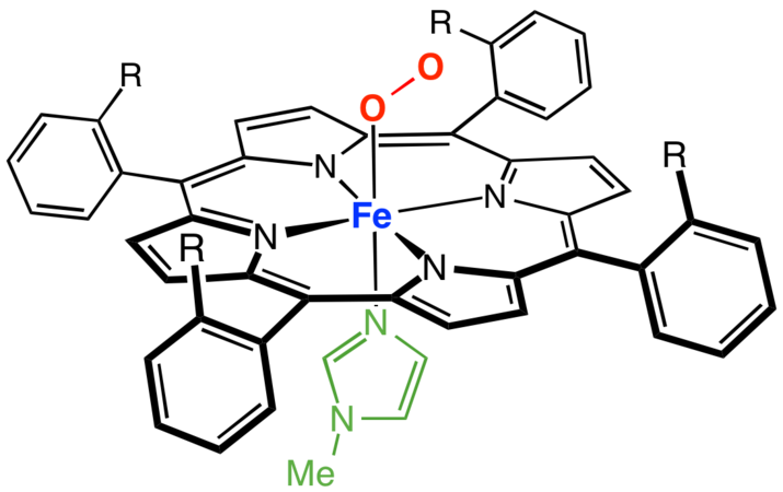
Myoglobin is a small monomeric protein, meaning it has only one polypeptide chain and one heme group, so it shows a well‑defined tertiary structure but no quaternary structure. Because of its high affinity for oxygen, myoglobin acts as the main oxygen storage protein in muscle, while hemoglobin transports oxygen in the blood.
Definition: Myoglobin is a muscle protein that binds and stores oxygen, ensuring a steady supply of oxygen to working muscles.
Basic Facts about MyoGlobin
These quick facts help answer many exam and viva questions.
- Symbol / name: Myoglobin, myoglobin, symbol Mb or MB.
- Location: Present mainly in cardiac muscle and red (slow‑twitch) skeletal muscle fibers of vertebrates, including humans.
- Type of protein: Monomeric globular protein with a compact tertiary structure and no quaternary structure.
- Number of amino acids: The human myoglobin chain contains about 153 amino acid residues.
- Molecular weight (molecular mass): Approximately 16.7 kDa (16,700 Da).
- Prosthetic group: One heme (iron–porphyrin) group per molecule, which binds one molecule of oxygen reversibly.
In one line: MyoGlobin is a monomeric heme protein in muscle that stores oxygen and has about 153 amino acids and a molecular weight near 16.7 kDa.
MyoGlobin Structure
Myoglobin is one of the best-studied proteins in biochemistry and is a classic example for understanding primary, secondary, and tertiary structure.

Primary structure of MyoGlobin
The primary structure of myoglobin is a single polypeptide chain of around 153 amino acids arranged in a specific sequence. This amino acid sequence determines how the chain folds to create a hydrophobic interior pocket to hold the heme group.
Key points:
- One polypeptide chain (monomer).
- About 153 amino acids in human myoglobin.
- The sequence forms the globin chain that surrounds the heme.
Secondary structure of MyoGlobin
The secondary structure of myoglobin is dominated by α‑helices. The chain folds into 8 major alpha helices, labeled A–H, which are connected by short non‑helical loops or turns.
Important features:
- High α‑helix content, very little or no β‑sheet.
- Helices pack together to form a compact “globular” shape.
- This pattern makes MyoGlobin a standard example of an α‑helical globular protein.
Student‑level statement:
MyoGlobin’s secondary structure is almost completely alpha‑helical, so it is often used as an example of α‑helix‑rich globular proteins.
Tertiary structure and heme group (active site)
The tertiary structure of MyoGlobin is a compact, globular 3‑D fold where the eight α‑helices pack around a central hydrophobic pocket. In this pocket lies the heme group, which is the active site of oxygen binding.
- The heme group consists of a protoporphyrin IX ring with a central ferrous iron ion (Fe²⁺).
- The Fe²⁺ is coordinated to four nitrogen atoms of the porphyrin ring and to one histidine residue (proximal His) from the globin chain; the sixth position binds oxygen.
- A distal histidine on the opposite side of heme stabilizes bound O₂ and reduces oxidation of iron to Fe³⁺.
Concept clarification:
Because MyoGlobin has only one polypeptide chain, it shows tertiary structure but no quaternary structure, unlike hemoglobin, which is a tetramer.
Functions of MyoGlobin in Muscle
The primary function of MyoGlobin is to store oxygen in muscle cells and release it when oxygen demand increases. It helps maintain aerobic metabolism in contracting muscles by acting as both an oxygen reservoir and a facilitator of oxygen diffusion.
Major functions:
- Oxygen storage: MyoGlobin binds oxygen strongly and stores it in muscle fibers for use when blood oxygen supply temporarily decreases, for example, during intense exercise.
- Oxygen transport within the muscle cell: It helps move oxygen from the sarcolemma and nearby capillaries to mitochondria located deeper inside the muscle cell.
- Buffering of intracellular oxygen: MyoGlobin desaturates at the onset of muscle activity, increasing the oxygen gradient from capillaries to mitochondria and smoothing fluctuations in oxygen concentration.
- Additional roles: MyoGlobin can participate in nitric oxide metabolism and protection against reactive oxygen and nitrogen species in muscle.
Easy to remember line:
In simple words, MyoGlobin is the oxygen storage protein of muscle: it stores oxygen, releases it when needed, and helps muscles keep working aerobically.
MyoGlobin vs Hemoglobin (Structure and Function)
MyoGlobin and hemoglobin are both heme proteins, but they differ in structure, location, and physiological role.
Structural differences
| Feature | MyoGlobin | Hemoglobin |
|---|---|---|
| Subunits | 1 polypeptide chain (monomer) | 4 polypeptide chains (tetramer: 2α + 2β) |
| Structure level | Tertiary only, no quaternary structure | Quaternary structure with cooperative subunits |
| Heme groups | 1 heme group per molecule | 4 heme groups per molecule |
| Location | Cardiac and skeletal muscle cells | Red blood cells (RBCs) |
| O₂‑binding curve | Hyperbolic; no cooperativity | Sigmoidal; strong cooperativity |
| O₂ affinity | High; releases O₂ at low pO₂ in muscle | Moderate; adapted for loading in lungs and unloading in tissues |
Functional differences
- MyoGlobin stores oxygen inside muscle cells and facilitates local oxygen delivery to mitochondria during contraction.
- Hemoglobin transports oxygen from lungs to tissues and returns carbon dioxide from tissues to lungs via the bloodstream.
- MyoGlobin’s high oxygen affinity and non‑cooperative binding make it ideal for oxygen storage, while hemoglobin’s cooperative binding makes it ideal for oxygen transport.
Key exam sentence:
MyoGlobin is a monomeric oxygen storage protein in muscle, whereas hemoglobin is a tetrameric oxygen transport protein in red blood cells.
Clinical Importance of MyoGlobin
MyoGlobin is clinically important because it is released into the bloodstream and urine when muscle cells are damaged. Raised myoglobin levels can signal acute myocardial infarction (heart attack) or severe skeletal muscle injury such as rhabdomyolysis.
Myoglobin as a diagnostic marker
- Serum myoglobin test: Blood myoglobin may rise within 1–3 hours after muscle injury and returns to normal within about 24–36 hours.
- Myoglobin is a sensitive but nonspecific marker of muscle damage because it is present in both heart and skeletal muscle.
- For myocardial infarction, more specific markers like cardiac troponins and CK‑MB are now preferred, but myoglobin can still be used as an early indicator.
Myoglobinuria and kidney damage
- Myoglobinuria refers to the presence of myoglobin in urine, often causing dark or “cola‑colored” urine in conditions with massive muscle breakdown.
- High levels of myoglobin in urine can be toxic to renal tubules and may lead to acute kidney injury if not treated promptly with adequate hydration and management.
MyoGlobin in Different Species and Adaptations
MyoGlobin is found in the striated muscles of almost all vertebrates and in many invertebrates. Its concentration varies widely and correlates with an animal’s dependence on sustained aerobic activity.
- Diving mammals such as whales and seals have extremely high MyoGlobin concentrations in skeletal muscles, enabling them to store large amounts of oxygen and remain underwater for long periods.
- Human MyoGlobin is encoded by the MB gene and is especially abundant in cardiac muscle and oxidative skeletal muscle fibers, supporting continuous aerobic work.
Frequently Asked Questions (Myoglobin FAQs)
What is MyoGlobin in simple terms?
MyoGlobin is an iron‑containing globular protein found in cardiac and skeletal muscle cells that binds and stores oxygen. It acts as the primary oxygen storage molecule inside muscle fibers, releasing oxygen when the muscle is actively contracting and the local oxygen level drops. MyoGlobin is a monomeric protein with a single polypeptide chain and one heme group
Where is MyoGlobin found?
MyoGlobin is found mainly in cardiac muscle and red slow‑twitch skeletal muscle fibers of vertebrates, including humans. It is located inside the muscle cell cytoplasm, where it stores oxygen close to the mitochondria. Normally, only very small amounts of myoglobin are present in the blood; elevated blood myoglobin levels indicate muscle injury or damage.
What is the primary function of MyoGlobin?
The primary function of MyoGlobin is to store oxygen inside muscle cells and release it when oxygen demand rises, such as during intense exercise. It also facilitates the diffusion of oxygen from capillaries to mitochondria within the muscle fiber. Without myoglobin, muscle cells would struggle to maintain aerobic metabolism during periods of high activity or reduced blood flow.
Is MyoGlobin an enzyme?
No, MyoGlobin is not an enzyme. It is a globular heme protein whose role is reversible oxygen binding and storage, not catalysis. Enzymes speed up chemical reactions, whereas MyoGlobin simply binds oxygen through its heme group and releases it when needed. MyoGlobin belongs to the globin protein family, similar to hemoglobin, not to the enzyme class.
Is MyoGlobin a globular or fibrous protein?
MyoGlobin is a globular protein. Its polypeptide chain folds into a compact, rounded three‑dimensional shape stabilized by hydrophobic interactions around the central heme group. The secondary structure is almost entirely made up of α‑helices, which pack together tightly to form the globular fold. This makes MyoGlobin a classic textbook example of a globular, α‑helical protein.
Does MyoGlobin have a quaternary structure?
No, MyoGlobin does not have a quaternary structure. Quaternary structure occurs only when two or more polypeptide chains associate together, as in hemoglobin, which has four subunits. MyoGlobin consists of just one polypeptide chain, so it only has primary, secondary, and tertiary structures. It is therefore described as a monomeric protein with tertiary structure only.
How many amino acids and what is the molecular weight of myoglobin?
Human MyoGlobin contains approximately 153 amino acid residues in its single polypeptide chain. Its molecular weight is approximately 16.7 kDa (16,700 Daltons), making it a relatively small protein. The molar mass of MyoGlobin is often cited as around 17 kDa in biochemistry exams. These values make MyoGlobin one of the most studied small heme proteins in molecular biology.
Which protein stores oxygen in muscle cells?
MyoGlobin is the protein that stores oxygen in muscle cells. It is an iron‑containing globular heme protein present in cardiac and skeletal muscle fibers. MyoGlobin binds oxygen with high affinity and releases it when the intracellular oxygen tension falls during muscle contraction. It acts as an intracellular oxygen reservoir, distinct from hemoglobin, which transports oxygen through the bloodstream.
What is the difference between MyoGlobin and hemoglobin?
MyoGlobin is a monomeric protein with one polypeptide chain and one heme group found in muscle, while hemoglobin is a tetrameric protein with four polypeptide chains and four heme groups found in red blood cells. MyoGlobin stores oxygen in muscles with high affinity and a hyperbolic binding curve, whereas hemoglobin transports oxygen in blood with cooperative binding and a sigmoidal curve.
Summary of Myoglobin
MyoGlobin is an iron‑containing, oxygen‑binding globular protein found in human cardiac and skeletal muscle, where it acts as the primary oxygen storage molecule.
Its structure consists of a single polypeptide chain (primary structure) rich in α‑helices (secondary structure) folded into a compact tertiary structure around a heme group that binds oxygen.
Because MyoGlobin has only one polypeptide chain, it has no quaternary structure and serves as a clear example of the tertiary structure of protein, often contrasted with the tetrameric hemoglobin.
Functionally, MyoGlobin stores oxygen in muscle, facilitates oxygen diffusion to mitochondria, and supports continuous aerobic metabolism, which is why questions like “what is the primary function of myoglobin” and “myoglobin stores oxygen in muscle cells” are common in exams.
Clinically, elevated blood or urine myoglobin can indicate muscle injury, and comparison topics such as “myoglobin vs hemoglobin,” “myoglobin structure and function”, and “hemoglobin and myoglobin structure and function” help students link its molecular features to its role in the body.
Discover more from Biochemistry Den
Subscribe to get the latest posts sent to your email.