In biochemistry, eicosanoids (preferred IUPAC name icosanoids) are signaling molecules made by oxidation of 20-carbon fatty acids. They exert complex control over many bodily systems such as
- In growth during and after physical activity,
- Inflammation or immunity after the intake of toxic compounds and pathogens, and
- act as messengers in the central nervous system.
The networks of controls that depend upon eicosanoids are among the most complex in the human body.
The first step in the formation of eicosanoid mediators consists in the release of the precursor fatty acids from the membrane phospholipids. This release may happen along several possible metabolic routes (Figure 1.1-1a).
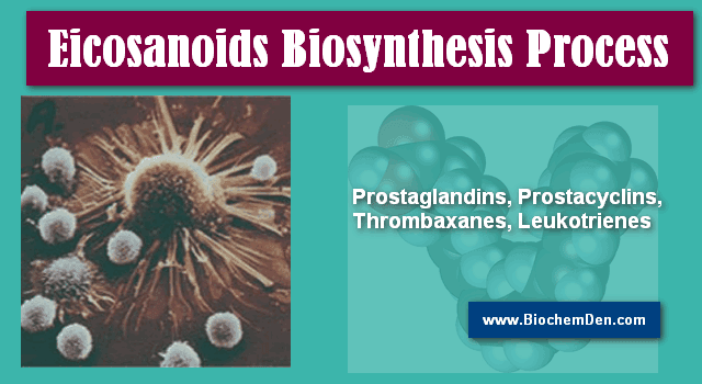
How Eicosanoids are synthesizing in Human body:
Figure 1.1-1:
a) Alternate pathways of Arachidonic acid release
b) cellular locations of enzymes involved in Eicosanoid formation
- a: Arachidonic acid may be directly released by phospholipase A2 (PLA2), or alternatively by the successive action of phospholipase C (PLC) and diacylglycerol (DAG) lipase.
- b: The major mechanism of release involves a cytosolic phospholipase A2 (cPLA2). An increase of Ca++ in response to an extrinsic signal causes binding of cPLA2 to the nuclear membrane. Cyclooxygenase (COX) and Lipoxygenase (LOX) form their respective intermediates, which are further processed by cytosolic enzymes to prostaglandins (PG), thromboxanes (TG), and leukotrienes (LT), respectively.
The major physiological mechanism of release consists in the activation of a Cytosolic phospholipase A2 (cPLA2) by Ca++ in response to an extracellular signal. cPLA2 then attaches to the nuclear (and probably ER) membranes, which appear to be the major reservoir of Arachidonic acid and its analogs.
- What are Lipids ? What are the Importance in Human Body
- Lipid Metabolism: Simple Smart Guide and Notes
The formation of the most important Eicosanoid derivatives of Arachidonic acid and its analogs is initiated by cyclooxygenases (Cox) and lipoxygenases (Lox):
- Cyclooxygenase, also called prostaglandin H synthase, converts arachidonic acid first into prostaglandin G2 (PGG2) and then PGH2. PGH2 is the common precursor of prostaglandins E2 and F2, and of prostaglandin I2 (prostacyclin). It is also the precursor of thromboxane A2 (TXA2). Therefore, cyclooxygenase is the single most important enzyme and drug target in eicosanoid metabolism.
- Lipoxygenase 5 converts arachidonic acid into 5-hydroperoxy-eicosatetraenoic acid (5-HPETE), which is the precursor of leukotrienes. Leukotrienes are formed mainly in leukocytes, e.g. macrophages and granulocytes, and they are potent pro-inflammatory mediators. Suppression of leukotriene synthesis with inhibitors of lipoxygenases is a fairly recent therapeutic principle in the treatment of asthma and chronic inflammation.
- 12- and 15-HPETE are formed by the corresponding lipoxygenases 12 and 15. They can be reduced to hydroxy- eicosatetraenoic acids (HETEs) and further converted to lipoxins (cf Figure 1.2-5). H have their own receptors and physiological roles, but they are not presently in the focus of interest of drug therapy or development.
Figure 1.2-5:

- Eicosanoids derived by lipoxygenases. a: Mechanism of reaction, illustrated for 15-lipoxygenase.
- Abstraction of a hydrogen atom by a non-heme iron in the active site leads to a carbon radical intermediate, which subsequently reacts with an oxygen π radical.
- b: Products of the three lipoxygenases (5-, 12-, and 15-lipoxygenase).
- The initial hydroperoxy products (HPETE = hydroperoxyeicosatetraenoic acid) are reduced by glutathione peroxidases to hydroxy derivatives (HETE). HETEs may have signalling roles of their own, and they may also be converted further to lipoxins.
- This conversion is initiated by another lipoxygenase, as shown here for 5-lipoxygenase (5-Lox) acting on 15-HETE. 5-HPETE is also the precursor of the leukotrienes.
The major ‘classical’ drug target in prostaglandin metabolism is cyclooxygenase, which occurs in several isoforms: Cox-1, Cox-2, and apparently in some mammals Cox-3. This is due to the central role of its product prostaglandin H2 as a precursor of multiple eicosanoid mediators (Figure 1.1-3).
Figure 1.1-3:

Structures of prostaglandin H2 and its derivatives. PG, prostaglandin; TX, thromboxane.
The synthesis of PGH2 occurs in two separate successive reactions (Figure 1.1-2), for which there are two separate active sites on the cyclooxygenase molecule.
- Essential Fatty Acids Definition and Notes in Biology
- Fatty Acid Synthesis: Activation, Steps and Control
Figure 1.1-2:

The two reactions catalyzed by cyclooxygenase. For each of these reactions, there is a separate active site.
The first reaction – also referred to as the cycloxygenase reaction – introduces two peroxy groups, one endoperoxide (forming a ring with carbons 9-11) and a hydroperoxide attached to position 15.
The resulting product– prostaglandin G2– is quite unstable, yet it is able to dissociate from the enzyme and to rebind to the second active site of the same or another enzyme molecule. There, the hydroperoxide is reduced to a simple hydroxy group, at the expense of two equivalents of reduced glutathione. This second step is called the peroxidase reaction.
Figure 1.1-4:

Spatial relationship of the two active sites in cyclooxygenase 1.
- a: Heme (green, with iron in magenta) and arachidonic acid (white, space-filling) bound within the peroxidase and the cyclooxygenase sites, respectively.
- b: Location of three important amino acid residues within the cyclooxgenase active site. Note that Tyr 385 is positioned between the two active sites.
- c: Space-filling representation of arachidonic acid (yellow), Tyr 385 (white), and heme (green).
The two active sites of cyclooxygenase are located close to each other (Figure 1.1-4a), and it is believed that this proximity is important in the ‘priming’ of the cyclooxygenase site. The first step in the cyclooxygenase reaction (Figure 1.1-5) is initiated by a tyrosyl radical (Tyr385 in cyclooxygenase 1; Figure 1.1-4b,c).
Figure 1.1-5:

- Catalytic mechanism of the cyclooxygenase reaction, leading from arachidonic acid (top left) to prostaglandin G2 (top right). Y· and YH represent Tyr 385.
- Molecular oxygen reacts in its π-radical form.
- Note that this is only the first one of the two reactions catalyzed by cyclooxygenase.
This tyrosyl radical will not exist in a newly translated enzyme molecule, and once it is there, it may be lost due to capture of a hydrogen from somewhere else than the substrate. There thus has to be a mechanism for its formation or regeneration. This mechanism is provided by the heme in the peroxidase active site.
- Hemoglobin: Structure, Function and its Properties
- Glycogenolysis : How Glycogen is Utilizing in Animals
The heme radical cation, which forms as an intermediate during the peroxidase reaction, can abstract a hydrogen atom from the tyrosine -OH group, which thus may act as a reductant in place of one of the glutathione molecules normally functioning as cosubstrates (Figure 1.1-6b).
Figure 1.1-6:

- Overview of the peroxidase reaction of cyclooxygenase. a: Prostaglandin G2 enters the peroxidase active site after leaving the cyclooxygenase site.
- It is reduced by heme, which thereby is converted into the cation radical form (red).
- The heme is reduced in turn by glutathion (GSH) in a stepwise fashion. b: ‘Priming’ of the cyclooxygenase site.
- The heme cation radical abstracts a hydrogen atom from Tyr 385, which thereby replaces one glutathione.
- The prostaglandin G (PGG) that is required to form the heme cation radical in the first place must be provided by another enzyme molecule. (AA-H: arachidonic acid)
The peroxidase reaction (Figure 1.1-6a) can function in the absence of cyclooxygenase activity, because the intermediate product (prostaglandin G2) can be provided by another enzyme molecule.
In accord with this model, a sample of cyclooxygenase, when expressed recombinantly and in the absence of arachidonic acid substrate, will initially be inactive, but it will exhibit ‘burst’ kinetics upon first contact with arachidonic acid, due to the cascading activation of more and more enzyme molecules by PGG2.