The mitosis occurs in the somatic cells and is meant for the multiplication of cell numbers during embryo-genesis and blastogenesis of plants and animals. Fundamentally, It remains related to the growth of an individual from zygote to adult stage. One of the basic characteristics of the mitotic cell division which is meant for the growth due to multiplication is that it gives rise to 2 daughter cells, which resembles each other and also the parent cell quantitatively and qualitatively. This basic outline remains the same in all living organisms.
Mitosis Stages:
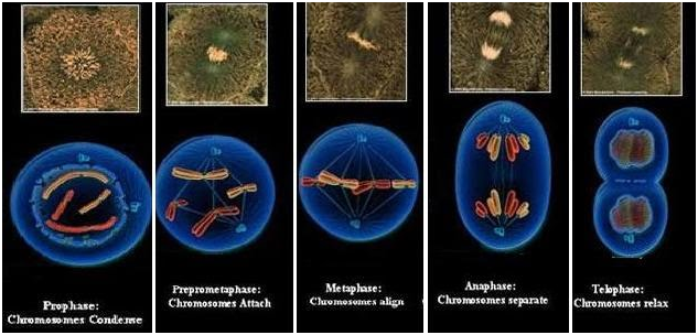
There are 5 stages in which the mitosis phase occurs they are
- Prophase
- Prometaphase
- Metaphase
- Anaphase
- Telophase
Let us see the mitosis stages in detail.
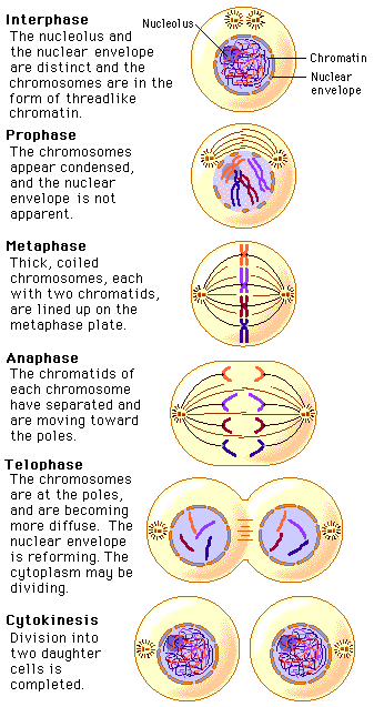
Stage I: Prophase
- During the prophase in the nucleus, nucleoli disappear and chromatin fibers condense into discrete, observable chromosomes, composed of two identical sister chromatids joined at the centromere.
- In the cytoplasm, Mitotic spindle starts to forms and is composed of microtubules between the two centrosomes or microtubule-organizing centers.
- During prophase centrosomes move apart, apparently propelled along the nuclear surface by lengthening of the microtubule bundles between them.
- In prophase, the nuclear membrane disappears.
In an animal cell, the centrioles move to the opposite ends/poles of the cell.
Stage II: prometaphase
- During prometaphase the nuclear envelope fragments, which allows microtubules to interact with the highly condensed chromosomes.
- During this phase, spindle fibers extend from each pole toward the equator of the cell.
- Each chromatid forms a specialized structure, the kinetochore, located at the centromere region.
- Microtubules called kinetochore microtubules to become attached to the kinetochores and keep the chromosomes into agitated motion. Microtubules called non-kinetochore microtubules radiate from the centrosome toward the metaphase plate without attaching to chromosomes. Nonkinetochore microtubules radiating from one pole interacts with those from the opposite ends.
Stage III: metaphase
- During metaphase: Centrosomes are moved at opposite poles of the cell. Chromosomes move to the plane equidistant between the spindle poles called metaphase plate.
- Centromeres of the present in all chromosomes are aligned on the metaphase plate.
- The long axis of each chromosome is roughly arranged at a right angle to the spindle axis.
- Kinetochores of sister chromatids face opposite poles and identical chromatids are attached to kinetochore fibers radiating from opposite ends of the parent cell.
Stage IV: anaphase
- Anaphase is characterized by the movement of chromosomes.
- Anaphase begins when paired centromeres of each chromosome move apart.
- Sister chromatids divide apart into separate chromosomes and move towards opposite poles.
- As the kinetochore fibers are attached to the centromeres, the chromosomes move centromere first in a “V” shape.
- Microtubules shorten at the kinetochore end as chromosomes approach the poles.
- At the same time, the poles of the cell move farther apart, elongating the cell.
- The two poles have identical collections of chromosomes.
Stage V: Telophase
- During telophase, the nonkinetochore microtubules further elongate the cell.
- Two daughter nuclei begin to format the two poles.
- New nuclear membrane form around the chromosomes from pieces of the parent cell’s nuclear envelope and portions of the endomembrane system.
- New nucleoli begin to reappear.
- Chromatin fiber uncoils and the chromosomes become less distinct.
- By the end of telophase, the equal division of one nucleus into two genetically identical nuclei is complete.